A cataract is a clouding of the lens inside the eye which leads to a decrease in vision. It is the most common cause of blindness and is conventionally treated with surgery. Visual loss occurs because opacification of the lens obstructs light from passing and being focused
on the retina at the back of the eye.
Cataracts, a leading cause of blindness among adults 45 years of age and older, are a clouding of the eye’s lens. Because of this cloudiness, light rays do not easily pass through the lens to focus on the retina. As a result, things look foggy or cloudy.
Most cataracts can be diagnosed with an eye exam. Your eye doctor will test your vision and examine your eyes with a slit lamp microscope to look for problems with the lens and other parts of the eye. The pupils are dilated to better examine the back of the eye, where the retina and optic nerve lie.
A nuclear cataract is the most common type of cataract, beginning with a gradual hardening and yellowing of the central zone of the lens, also known as the nucleus. Over time, this hardening and yellowing will expand to the other layers of the lens.
Cataracts are cloudy areas in the lens of the eye that can cause changes in vision. Symptoms of cataracts include cloudy or fuzzy vision as well as sensitivity to glare. Cataracts are treated with surgery.
What Are Cataracts?
Cataract is a clouding of the eye’s lens. When we look at something, light rays travel into our eye through the pupil and are focused through the lens onto the retina, a layer of light-sensitive cells at the back of the eye. The lens must be clear in order to focus light properly onto the retina. If the lens has become cloudy, this is called a cataract.
Vision problems with cataracts
If your vision has become blurry, cloudy or dim, or things you see are not as bright or colorful as they used to be, a cataract may have developed in one or both of your eyes. Many people say that their vision with cataracts is similar to the effect of looking through a dirty car windshield.
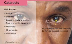
Cataracts are most commonly due to aging, but may also occur due to trauma, radiation exposure, be present from birth, or occur following eye surgery for other problems.[1][6] Risk factors include diabetes, smoking tobacco, prolonged exposure to sunlight, and alcohol. Either clumps of protein or yellow-brown pigment may be deposited in the lens reducing the transmission of light to the retina at the back of the eye. Diagnosis is by an eye examination.[1]
Prevention includes wearing sunglasses and not smoking. Early on the symptoms may be improved with eyeglasses. If this does not help, surgery to remove the cloudy lens and replace it with an artificial lens is the only effective treatment. Surgery is only needed if the cataracts are causing problems.[1] Surgery generally results in an improved quality of life.[7] Cataract surgery is not easily available in many countries, which is especially true for women, those living in rural areas, and those who cannot read.[6][8]
Signs and symptoms[edit]
Signs and symptoms vary depending on the type of cataract, though considerable overlap occurs. People with nuclear sclerotic or brunescent cataracts often notice a reduction of vision. Those with posterior subcapsular cataracts usually complain of glareas their major symptom.[10]
The severity of cataract formation, assuming no other eye disease is present, is judged primarily by a visual acuity test. The appropriateness of surgery depends on a patient’s particular functional and visual needs and other risk factors, all of which may vary widely.[11]
Causes
Age is the most common cause.[1][6] Lens proteins denature and degrade over time, and this process is accelerated by diseases such as diabetes mellitus and hypertension. Environmental factors, including toxins, radiation, and ultraviolet light, have cumulative effects, which are worsened by the loss of protective and restorative mechanisms due to alterations in gene expression and chemical processes within the eye.[12]
Blunt trauma causes swelling, thickening, and whitening of the lens fibers. While the swelling normally resolves with time, the white color may remain. In severe blunt trauma, or injuries which penetrate the eye, the capsule in which the lens sits can be damaged. This allows fluid from other parts of the eye to rapidly enter the lens leading to swelling and then whitening, obstructing light from reaching the retina at the back of the eye. Cataracts may develop in 0.7 to 8.0% of cases following electrical injuries.[13]
Ultraviolet light, specifically UVB, has been shown to cause cataracts, and some evidence indicates sunglasses worn at an early age can slow its development in later life.[14] Microwave radiation has also been found to cause cataracts. The mechanism is unclear, but it may include changes in heat-sensitive enzymes that normally protect cell proteins in the lens. Another possible mechanism is direct damage to the lens from pressure waves induced in the aqueous humor.
Cataracts have been associated with ionizing radiation such as X-rays. The addition of damage to the DNA of the lens cells has been considered.[15] Finally, electric and heat injuries denature and whiten the lens as a result of direct protein coagulation.[12] This same process makes the clear albumin of an egg become white and opaque after cooking. Cataracts of this type are often seen in glassblowersand furnace workers. Lasers of sufficient power output are known to damage the eyes and skin.
Genetics
The genetic component is strong in the development of cataracts,[16] most commonly through mechanisms that protect and maintain the lens. The presence of cataracts in childhood or early life can occasionally be due to a particular syndrome. Examples of chromosome abnormalities associated with cataracts include 1q21.1 deletion syndrome, cri-du-chat syndrome, Down syndrome, Patau’s syndrome, trisomy 18 (Edward’s syndrome), and Turner’s syndrome, and in the case of neurofibromatosis type 2, juvenile cataract on one or both sides may be noted. Examples of single-gene disorderinclude Alport’s syndrome, Conradi’s syndrome, myotonic dystrophy, and oculocerebrorenal syndrome or Lowe syndrome.
The skin and the lens have the same embryological origin and so can be affected by similar diseases.[17] Those with atopic dermatitis and eczema occasionally develop shield ulcers cataracts. Ichthyosisis an autosomal recessive disorder associated with cuneiform cataracts and nuclear sclerosis. Basal-cell nevus and pemphigus have similar associations.
Cigarette smoking has been shown to double the rate of nuclear sclerotic cataracts and triple the rate of posterior subcapsular cataracts.[18] Evidence is conflicting over the effect of alcohol. Some surveys have shown a link, but others which followed patients over longer terms have not.[19]
Medications
Some drugs, such as corticosteroids, can induce cataract development.[20] People with schizophrenia often have risk factors for lens opacities (such as diabetes, hypertension, and poor nutrition) butantipsychotic medications are unlikely to contribute to cataract formation.[21] Miotics[22] and triparanol may increase the risk.[23]
Healthcare caused
Nearly every person who undergoes a vitrectomy — without ever having had cataract surgery — will experience progression of nuclear sclerosis at 6-months and 12-month after the operation.[24] This may be because the native vitreous humor is significantly different to the solutions used to replace the vitreous (vitreous substitutes), such as BSS Plus.[25] This may also be because the native vitreous humour contains ascorbic acid which helps neutralize oxidative damage to the lens and because traditional vitreous substitutes do not contain ascorbic acid.[26][27] As such, for phakic patients requiring a vitrectomy it is becoming increasingly common for ophthalmologists to offer the vitrectomy with a combined prophylactic cataract surgery procedure to prophylactically prevent cataract formation.[28]
Other diseases
· Metabolic and nutrition diseases · Aminoaciduria or Lowe’s syndrome · Galactosemia / galactosemic cataract · Cytomegalic inclusion disease · Rubella | · Genetic syndromes · Infections: · Leprosy · Secondary to other eye diseases: · Aniridia · Uveitis |
Classification
Cross-sectional view, showing the position of the human lens
Cataracts may be partial or complete, stationary or progressive, or hard or soft. The main types of age-related cataracts are nuclear sclerosis, cortical, and posterior subcapsular.
Nuclear sclerosis, the most common type of cataract, involves the central or ‘nuclear’ part of the lens. Over time, this becomes hard or ‘sclerotic’ due to condensation of lens nucleus and deposition of brown pigment within the lens. In advanced stages, it is called brunescent cataract. This type of cataract can present with a shift to nearsightedness and causes problems with distance vision, while reading is less affected.[29]
Cortical cataracts are due to the lens cortex (outer layer) becoming opaque. They occur when changes in the fluid contained in the periphery of the lens causes fissuring. When these cataracts are viewed through an ophthalmoscope or other magnification system, the appearance is similar to white spokes of a wheel. Symptoms often include problems with glare and light scatter at night.[29]
Posterior subcapsular cataracts are cloudy at back of the lens adjacent to the capsule (or bag) in which the lens sits. Because light becomes more focused toward the back of the lens, they can cause disproportionate symptoms for their size.
An immature cataract has some transparent protein, but with a mature cataract, all the lens protein is opaque. In a hypermature or Morgagnian cataract, the lens proteins have become liquid. Congenital cataract, which may be detected in adulthood, has a different classification and includes lamellar, polar, and sutural cataracts.[20][30]
Cataracts can be classified by using the lens opacities classification system LOCS III. In this system, cataracts are classified based on type as nuclear, cortical, or posterior. The cataracts are further classified based on severity on a scale from 1 to 5. The LOCS III system is highly reproducible.[31]
Cataract symptom progression
See a simulation of what vision with cataract looks like.Get a baseline exam at age 40 when early signs of disease and changes in vision may start to occur. Your ophthalmologist (Eye M.D.) will let you know how often you should return for follow-up exams. At any point, if you have symptoms or risks for eye disease, see your Eye M.D. Because your risk for cataracts and other eye diseases increases as you get older, starting at age 65 you should see your Eye M.D. every year. A complete eye examination will rule out any other condition that may be causing blurred vision or eye problems. Early detection and treatment of cataracts is critical to preserving sight.Cataracts happen when protein builds up in the lens of your eye, making it cloudy. This prevents light from passing clearly through the lens, causing some loss of vision. New lens cells form on the outside of the lens, and the older cells are compacted into the center of the lens, forming the cataract.
Other things that can raise your risk of getting cataracts include cigarette smoke, air pollution, and heavy drinking.
Cataract Symptoms and Signs
Cataracts are the most common cause of vision loss in people over age 40 and is the principal cause of blindness in the world. In fact, there are more cases of cataracts worldwide than there are ofglaucoma, macular degeneration and diabetic retinopathy combined, according to Prevent Blindness America (PBA).
One theory of cataract formation that’s gaining favor is that many cataracts are caused by oxidative changes in the human lens. This is supported by nutrition studies that show fruits and vegetables high inantioxidants may help prevent certain types of cataracts (see below).
Risk factors such as UVB exposure and smoking can be addressed, but are unlikely to make a large difference to visual function. Although no means of preventing cataracts has been scientifically proven, wearing ultraviolet-protecting sunglasses may slow the development.[32][33] While regular intake of antioxidants (such as vitamins A, C, and E) has been thought to protect against the risk of cataracts, clinical trials have shown it does not.[34] Evidence is mixed, but weakly positive, for a potential protective effect of the nutrients lutein and zeaxanthin.[35] Statin use is somewhat associated with a lower risk of nuclear sclerotic cataract.[36]


Cataract removal can be performed at any stage and no longer requires ripening of the lens. Surgery is usually ‘outpatient’ and performed using local anesthesia. About 9 of 10 patients can achieve a corrected vision of 20/40 or better after surgery.[29]
Several recent evaluations found surgery can only meet expectations when significant functional impairment from poor vision exists prior to surgery. Visual function estimates such as VF-14 have been found to give more realistic estimates than visual acuity testing alone.[29][37] In some developed countries, a trend to overuse cataract surgery has been noted, which may lead to disappointing results.[38]
Phacoemulsification is the most widely used cataract surgery in the developed world.[39][40] This procedure uses ultrasonic energy to emulsify the cataract lens. Phacoemulsification typically comprises six steps:
Extracapsular cataract extraction (ECCE) consists of removing the lens manually, but leaving the majority of the capsule intact.[41] The lens is expressed through a 10- to 12-mm incision which is closed with sutures at the end of surgery. ECCE is less frequently performed than phacoemulsification, but can be useful when dealing with very hard cataracts or other situations where emulsification is problematic. Manual small incision cataract surgery (MSICS) has evolved from ECCE. In MSICS, the lens is removed through a self-sealing scleral tunnel wound in the sclera which, ideally, is watertight and does not require suturing. Although “small”, the incision is still markedly larger than the portal in phacoemulsion. This surgery is increasingly popular in the developing world where access to phacoemulsification is still limited.
Intracapsular cataract extraction (ICCE) is rarely performed.[42] The lens and surrounding capsule are removed in one piece through a large incision while pressure is applied to the vitreous membrane. The surgery has a high rate of complications.

The postoperative recovery period (after removing the cataract) is usually short. The patient is usually ambulatory on the day of surgery, but is advised to move cautiously and avoid straining or heavy lifting for about a month. The eye is usually patched on the day of surgery and use of an eye shield at night is often suggested for several days after surgery.[11]
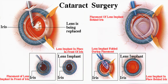
In all types of surgery, the cataractous lens is removed and replaced with an artificial lens, known as an intraocular lens, which stays in the eye permanently. Intraocular lenses are usually monofocal, correcting for either distance or near vision. Multifocal lenses may be implanted to improve near and distance vision simultaneously, but these lenses may increase the chance of unsatisfactory vision.[12]
Serious complications of cataract surgery include retinal detachment and endophthalmitis.[43] In both cases, patients notice a sudden decrease in vision. In endophthalmitis, patients often describe pain. Retinal detachment frequently presents with unilateral visual field defects, blurring of vision, flashes of light, or floating spots.
The risk of retinal detachment was estimated as about 0.4% within 5.5 years, corresponding to a 2.3-fold risk increase compared to naturally expected incidence, with older studies reporting a substantially higher risk. The incidence is increasing over time in a somewhat linear manner, and the risk increase lasts for at least 20 years after the procedure. Particular risk factors are younger age, male sex, longer axial length, and complications during surgery. In the highest risk group of patients, the incidence of pseudophakic retinal detachment may be as high as 20%.[44][45]
The risk of endophthalmitis occurring after surgery is less than one in 1000.[46]
Corneal edema and cystoid macular edema are less serious but more common, and occur because of persistent swelling at the front of the eye in corneal edema or back of the eye in cystoid macular edema.[47] They are normally the result of excessive inflammation following surgery, and in both cases, patients may notice blurred, foggy vision. They normally improve with time and with application of anti-inflammatory drops. The risk of either occurring is around one in 100.
Posterior capsular opacification, also known as after-cataract, is a condition in which months or years after successful cataract surgery, vision deteriorates or problems with glare and light scattering recur, usually due to thickening of the back or posterior capsule surrounding the implanted lens, so-called ‘posterior lens capsule opacification’. Growth of natural lens cells remaining after the natural lens was removed may be the cause, and the younger the patient, the greater the chance of this occurring. Management involves cutting a small, circular area in the posterior capsule with targeted beams of energy from a laser, called Nd:YAG laser capsulotomy, after the type of laser used. The laser can be aimed very accurately, and the small part of the capsule which is cut falls harmlessly to the bottom of the inside of the eye. This procedure leaves sufficient capsule to hold the lens in place, but removes enough to allow light to pass directly through to the retina. Serious side effects are rare.[48]Posterior capsular opacification is common and occurs following up to one in four operations, but these rates are decreasing following the introduction of modern intraocular lenses together with a better understanding of the causes.
Vitreous touch syndrome is a possible complication of intracapsular cataract extraction.[49]
Though there is significant controversy about whether cataracts can be prevented, a number of studies suggest certain nutrients and nutritional supplements may reduce your risk of cataracts.
One large, 10-year study of female health professionals found that higher dietary intakes of vitamin E and the carotenoids lutein andzeaxanthin from food and supplements were associated with significantly decreased risks of cataract.
Good food sources of vitamin E include sunflower seeds, almonds and spinach. Good sources of lutein and zeaxanthin include spinach, kale and other green, leafy vegetables.
Other studies have shown antioxidant vitamins such as vitamin C and foods containing omega-3 fatty acids may reduce cataract risk.
Visit our Nutrition & Eyes section to read more about eye vitaminsand how a healthful diet and good nutrition may help prevent cataracts.
Another step you can take to reduce your risk of cataracts is to wear protective sunglasses that block 100 percent of the sun’s UV rays when you are outdoors.
When symptoms begin to appear, you may be able to improve your vision for a while using new glasses, strongbifocals, magnification, appropriate lighting or other visual aids.
Think about surgery when your cataracts have progressed enough to seriously impair your vision and affect your daily life. Many people consider poor vision an inevitable fact of aging, but cataract surgery is a simple, relatively painless procedure to regain vision.